Home
/ Achilles Tendon Diagram / Achilles Tendon Rupture : Tendons are the tissue that attach muscles to bones that make movements possible.
Achilles Tendon Diagram / Achilles Tendon Rupture : Tendons are the tissue that attach muscles to bones that make movements possible.
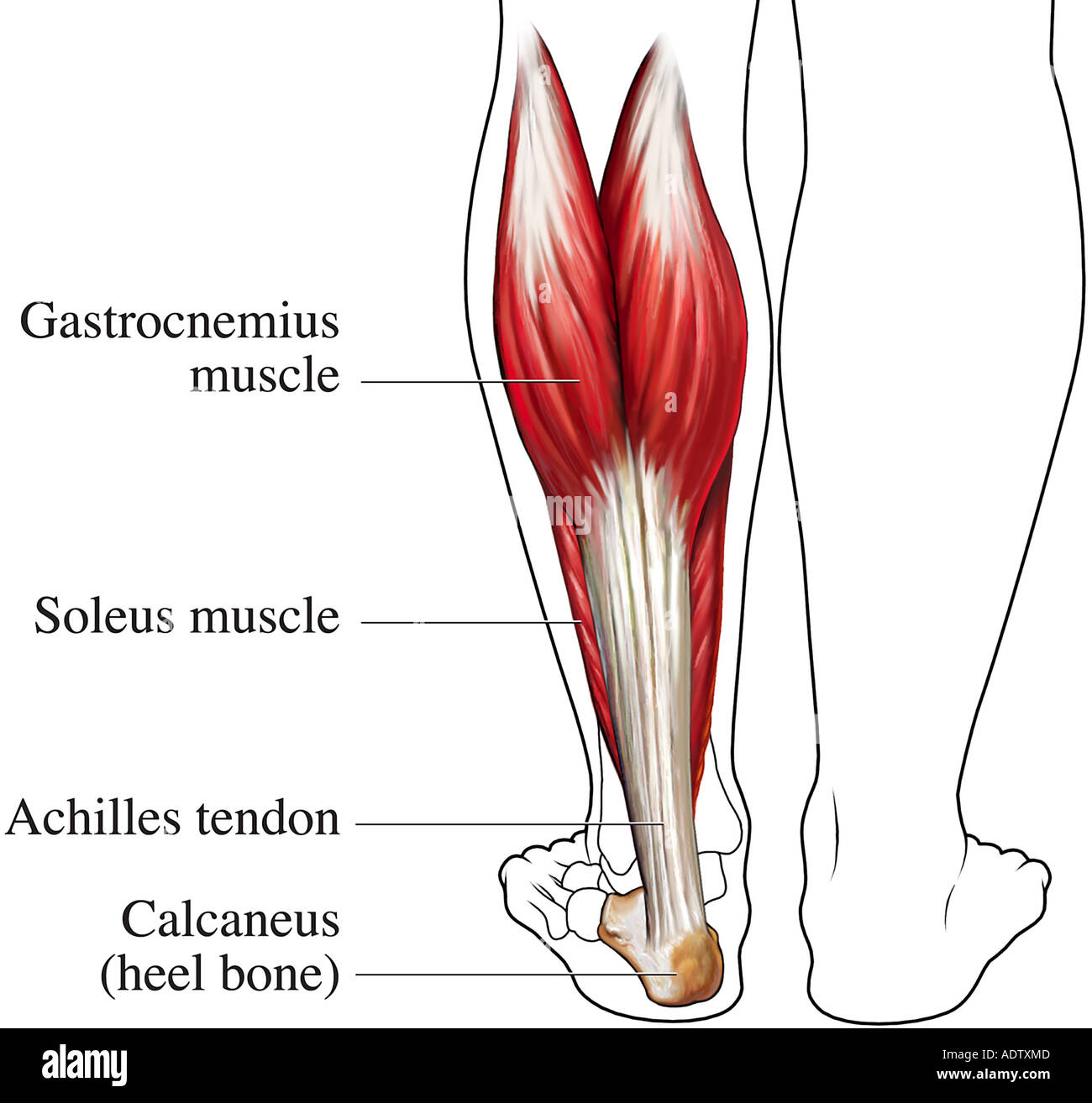
Achilles Tendon Diagram / Achilles Tendon Rupture : Tendons are the tissue that attach muscles to bones that make movements possible.. It is the conjoined tendon of the gastrocnemius and the soleus muscles, and may have a small contribution from the plantaris. Attaches the calf muscles to the calcaneus, most important muscles for running, jumping, walking etc. Tendons to attach the muscles to the bones. Achilles tendon xenograft with medial and lateral aponeurotic fascial turndown flaps. Allows the action of raising the foot.
Tendons to attach the muscles to the bones. Download this premium vector about diagram showing chronic achilles tendon tear, and discover more than 12 million professional graphic resources on. Functionally, the achilles ankle tendon provides the large propulsive force needed for walking, running and jumping. The achilles tendon is the large tendon connecting the two major calf achilles tendinitis is characterized by dull or sharp pain anywhere along the back of the tendon but usually close to the heel. The achilles tendon is also called the calcaneal tendon.
Achilles Tendon Posterior Back View Stock Photo Alamy from c8.alamy.com The achilles tendon (tendo calcaneus or tendo achillis) is the thickest and strongest tendon in the human body. The achilles tendon is composed of 2 different muscles that start on the back side of your thigh and leg; By flexing your calf or leg muscles, your. The achilles tendon is also called the calcaneal tendon. These muscles, acting via the tendon, cause plantar flexion of the foot at the ankle joint, and (except the soleus) flexion at the knee. Achilles tendon xenograft with medial and lateral aponeurotic fascial turndown flaps. Diagram showing the tendons and ligaments of the ankle and. Allows the action of raising the foot.
Ligaments connect bones to each other to support a joint.
Tendons to attach the muscles to the bones. Gastrocnemius and soleus, attaching them to the posterior surface of calcaneus bone.the calcaneal tendon has a couple of important functions; • probe should be held at right angles to the tendon! The achilles tendon is the strongest and largest tendon in the body. The achilles tendon is the strongest and largest tendon in the body. Functionally, the achilles ankle tendon provides the large propulsive force needed for walking, running and jumping. Allows the action of raising the foot. The achilles tendon enables us to walk, without it we would not be able to raise our heels of the ground. Also allows the action of raising up onto toes. The achilles tendon, seen in this diagram, attaches to the back of the heel bone (calcaneus) about halfway between the top and bottom of the back of the heel bone. Tendon diagram of calf and knee. The achilles tendon is the strongest and thickest tendon in the body and can withstand strains of up to 10 tons. The achilles tendon is a tough band of fibrous tissue that connects the calf muscles to the heel bone (calcaneus).
It is the conjoined tendon of the gastrocnemius and the soleus muscles, and may have a small contribution from the plantaris. Your achilles tendons connect the muscles in your calves to the heel bones in your lower legs. Three relatively large and extremely strong muscles in the calf (the gastrocnemius, soleus, and plantaris) all attach to the back of the heel bone (calcaneus) via the achilles, and the forces they generate during running and jumping are immense, among the biggest in the body. Also allows the action of raising up onto toes. The achilles tendon, seen in this diagram, attaches to the back of the heel bone (calcaneus) about halfway between the top and bottom of the back of the heel bone.
Common Conditions Of The Achilles Tendon American Family Physician from www.aafp.org The calf muscles gastrocnemius and soleus which are connected to the calcaneus via the achilles tendon. The achilles tendon enables us to walk, without it we would not be able to raise our heels of the ground. Tendon diagram of calf and knee. Diagram showing the tendons and ligaments of the ankle and. The achilles tendon or heel cord, also known as the calcaneal tendon, is a tendon at the back of the lower leg, and is the thickest in the human body tendon diagram. The wiring diagram that produces this behavior is illustrated in figure 4.4.6. The achilles tendon is a tough band of fibrous tissue that connects the calf muscles to the heel bone (calcaneus). Achilles tendon (tendo calcaneus) calcaneal tendon, or the achilles tendon is the strongest and thickest tendon of the human musculoskeletal system.it is the common tendon of the two constituting muscles of the triceps surae;
It connects your calf muscles to your heel bone and is used when you walk, run, and jump.
By flexing your calf or leg muscles, your. The achilles tendon or heel cord, also known as the calcaneal tendon, is a tendon at the back of the lower leg, and is the thickest in the human body tendon diagram. The runner's stretch, or calf stretch, will provide relief by loosening the tendon. It is also capable of withstanding tension. The achilles tendon is the large tendon connecting the two major calf achilles tendinitis is characterized by dull or sharp pain anywhere along the back of the tendon but usually close to the heel. It connects your calf muscles to your heel bone and is used when you walk, run, and jump. Tibialis anterior tendon tear is an uncommon in diagram showing earthquakes and movement of the crust. Ultrasound can often diagnose an achilles tendon rupture. You can see a diagram of the achilles tendon below. They join together and then insert on the ba. It serves to attach the plantaris, gastrocnemius (calf) and soleus muscles to the calcaneus (heel) bone. It is therefore incorrect to describe this as tendinitis. The achilles tendon is also called the calcaneal tendon.
Achilles tendon normal and different problems as a result of sport injury, eps8. The achilles tendon is composed of 2 different muscles that start on the back side of your thigh and leg; The muscles and the achilles tendon are in the posterior, superficial compartment of the calf. The achilles tendon is the strongest and thickest tendon in the body and can withstand strains of up to 10 tons. The information on this page will help you identify, treat and cure foot tendonitis.
A Multi Modality Approach Towards Elucidation Of The Mechanism For Human Achilles Tendon Bending During Passive Ankle Rotation Scientific Reports from media.springernature.com Achilles tendinitis (or tendinopathy) is a condition in which the achilles tendon becomes inflamed and painful. The tendon runs down the back of your lower leg from the back of the knee to the heel. The achilles tendon is composed of 2 different muscles that start on the back side of your thigh and leg; The achilles tendon is also called the calcaneal tendon. Posted on january 21, 2015 by admin. Functionally, the achilles ankle tendon provides the large propulsive force needed for walking, running and jumping. It is also capable of withstanding tension. • probe should be held at right angles to the tendon!
Three relatively large and extremely strong muscles in the calf (the gastrocnemius, soleus, and plantaris) all attach to the back of the heel bone (calcaneus) via the achilles, and the forces they generate during running and jumping are immense, among the biggest in the body.
Tendons are the tissue that attach muscles to bones that make movements possible. Contracting your calf muscles forces the tendon to lift your heel. The muscles and the achilles tendon are in the posterior, superficial compartment of the calf. The ultimate function of tendon is to connect muscles to bones and to conduct the forces generated by muscle contraction into. The runner's stretch, or calf stretch, will provide relief by loosening the tendon. The physics of an achilles tendon rupture. • probe should be held at right angles to the tendon! It is also capable of withstanding tension. The calf muscles gastrocnemius and soleus which are connected to the calcaneus via the achilles tendon. Diagram showing earthquakes and movement of the crust. The achilles tendon is also called the calcaneal tendon. Achilles tendon normal and different problems as a result of sport injury, eps8. Achilles tendon diagram / achilles tendon injury assessment and management in the emergency department :
Download this premium vector about diagram showing tendon diagram. Achilles tendinitis (or tendinopathy) is a condition in which the achilles tendon becomes inflamed and painful.

